Paulo Lobo1*, Ryan Nishi2, Chloe McCreery2, Ruyan Zhang1, Justin H Wong2, Peter Deptula1,3
1Department of Surgery, John A. Burns School of Medicine, University of Hawaiʻi at Mānoa, Honolulu, HI, USA
2John A. Burns School of Medicine, University of Hawaiʻi at Mānoa, Honolulu, HI, USA
3The Queen’s Medical Center, Honolulu, HI, USA
*Corresponding Author: Paulo Lobo, Department of Surgery, John A. Burns School of Medicine, University of Hawaiʻi at Mānoa, Honolulu, HI, USA
Received Date: 20 September 2026
Accepted Date: 26 September 2026
Published Date: 28 September 2026
Citation: Lobo P, Nishi R, McCreery C, Zhang R, Wong JH, et al. (2026) Expanding the Hemisoleus Flap: Versatile Applications for Complex Lower Extremity Reconstruction. J Surg 11: 11696 DOI: 10.29011/2575-9760.11696
Abstract
Introduction: Soft-tissue defects of the middle and distal thirds of the leg represent a persistent reconstructive challenge. Free tissue transfer remains the gold standard approach; however, not all patients are suitable candidates due to comorbidities, compromised recipient vessels, or resource constraints. Hemisoleus muscle flaps offer a reliable alternative for achieving soft-tissue coverage and limb preservation. This case series aims to characterize the breadth of hemisoleus-based flap techniques and their reliability for coverage of complex lower extremity wounds.
Methods: A retrospective review was performed for patients who underwent hemisoleus flap reconstruction between September 2023 and December 2025 at a single Level I trauma center. All procedures were performed by fellowship-trained reconstructive surgeons. Patient demographics, reconstruction indications, flap configurations, adjunctive procedures, and outcomes were reviewed.
Results: Twelve patients underwent reconstruction with hemisoleus-based flaps. Indications included traumatic wounds (n=8), infection (n=3), and oncologic defects (n=1). Defect sizes ranged from approximately 50 to 600 cm², with muscle flaps used to provide vascularized coverage of critical structures within larger wounds as needed. Hemisoleus flaps were used alone (n=4) or combined with additional regional muscle flaps (n=8). Complete flap survival was achieved in all cases with little morbidity.
Conclusion: Hemisoleus muscle flaps are reliable options for reconstruction of middle and distal leg defects. Their versatility, dependable vascularity, and ability to be used in multiple configurations make them valuable options for limb salvage when free tissue transfer is undesirable or impractical.
Keywords: Hemisoleus Flap; Lower Extremity Reconstruction; Limb Salvage; Muscle Flap; Propeller Flap; Soft-Tissue Defects
Introduction
Soft-tissue defects of the middle and distal thirds of the leg remain a challenging reconstructive problem, particularly in the setting of exposed bone, tendon, hardware, or chronic infection. Although free tissue transfer is often considered the gold standard for complex lower extremity reconstruction, not all patients are appropriate candidates because of medical comorbidities, poor recipient vessel quality, prolonged operative time, limited microsurgical resources, or local traumatic injury patterns. In these situations, regional muscle flaps continue to provide reliable and durable options for limb salvage while avoiding the complexity and resource demands of free tissue transfer. Among local reconstructive options, the hemisoleus flap represents a versatile yet underutilized technique for coverage of middle- and distalthird tibial defects. The soleus muscle possesses independent medial and lateral vascular territories supplied by perforators from the posterior tibial and peroneal arterial systems, respectively, allowing safe longitudinal splitting and preservation of perfusion to each muscle belly. Early anatomic and clinical studies established the vascular basis of the hemisoleus flap and demonstrated reliable healing using distally based split-soleus and medial hemisoleus designs for lower extremity defects [1, 2]. Subsequent clinical series further validated the reliability of proximally based medial hemisoleus flaps for coverage of exposed tibia, tendon, and hardware with acceptable donor-site morbidity and preservation of ambulatory function [3,5]. Continued refinements in flap design have expanded the reconstructive utility of the hemisoleus muscle. Distally based (reverse-flow) medial hemisoleus flaps based on distal posterior tibial artery perforators allow reliable coverage of distal-third tibial defects that may be beyond the reach of traditional proximally based constructs [4,6,7]. Angiosome- and perforator-based modifications have further improved flap reliability and reduced rates of distal necrosis [8]. More recently, propeller hemisoleus techniques have been described in which the muscle is islanded on a reverse-flow posterior tibial pedicle and rotated up to 180 degrees to increase the arc of rotation and extend coverage into the distal leg and foot [9]. In addition, combined gastrocnemius-hemisoleus “double flap” constructs have been utilized for larger proximal- and middle-third tibial defects, allowing expanded pedicled coverage while preserving the advantages of local tissue reconstruction [10-14]. Despite these advances, contemporary literature regarding hemisoleus reconstruction remains fragmented, with most reports focusing on individual flap configurations rather than a cohesive framework for selecting and applying different hemisoleus-based techniques. Practical considerations regarding intraoperative decision-making, perforator selection, and integration of hemisoleus flaps into modern reconstructive algorithms are also incompletely defined. The purpose of this study is to present a case series demonstrating the versatility and reliability of hemisoleus muscle flaps for reconstruction of complex lower extremity defects. We describe distinct clinical applications of hemisoleus-based reconstruction, including traditional proximally based, reverse-flow, propeller, and double constructs, while highlighting relevant anatomy, operative technique, and clinical considerations that support the broader use of hemisoleus-based reconstruction.
Methods
A retrospective review was conducted of patients who underwent hemisoleus muscle flap reconstruction for lower extremity softtissue defects at a single Level I trauma center between September 2023 and December 2025. Institutional review board approval was obtained prior to data collection. Patients were included if they underwent reconstruction with any hemisoleus-based flap configuration for coverage of middle and distal leg defects. Collected variables included patient demographics (age, sex, and comorbidities), wound characteristics (etiology, size, location, and presence of exposed bone, tendon, or hardware), operative details (flap configuration, adjunctive procedures, and need for staged reconstruction), and postoperative outcomes. Complications were classified as minor (wound events not threatening flap viability, managed without reoperation) or major (requiring return to the operating room or resulting in total flap loss). Outcomes were assessed per patient rather than per individual muscle flap. Flap selection was based on defect location, size, and intraoperative assessment of perforator anatomy. Four distinct hemisoleusbased reconstructive approaches were employed: (1) proximally based hemisoleus flap, (2) reverse hemisoleus flap, (3) propeller hemisoleus flap, and (4) double hemisoleus constructs. Additional regional muscle flaps were used when needed to supplement coverage. Primary outcomes included flap survival and successful wound coverage. Secondary outcomes included complication rates and need for reoperation. Postoperative outcomes were abstracted from available clinical records; follow-up was not standardized. Complication-related reoperation was distinguished from planned staged reconstruction, including delayed skin grafting.
Operative Technique
All cases begin with thorough excisional debridement to viable, bleeding tissue. Wound size, location, and the presence of exposed bone, tendon, or hardware are assessed to guide flap selection. Perforator anatomy is evaluated using handheld Doppler for cases of distally based or perforator-based flaps. The operative limb is elevated for exsanguination and a tourniquet is inflated to 250 mmHg. A longitudinal incision is designed over the medial calf for flap harvest. In cases where the defect has partially exposed this area, exposure is extended distally and proximally in a longitudinal fashion. The medial gastrocnemius and medial soleus muscles are identified within the superficial posterior compartment. The interval between these muscles is developed, and the soleus is elevated off the deep posterior compartment. The plantaris tendon serves as a useful landmark to confirm the correct plane; however, tissue planes may be obscured by prior trauma, chronic inflammation, infection, or previous surgery. The medial hemisoleus is then dissected free from its attachments to the tibia and deep posterior compartment using a combination of blunt and sharp dissection. The posterior tibial neurovascular bundle is carefully protected throughout, as it lies anteriorly within the deep compartment. Perforators arising from the posterior tibial bundle are identified in this interval and preserved until the final reconstructive plan is confirmed. The hemisoleus is then elevated off the conjoint tendoachilles. We recommend incising the overlying fascia proximal to the well-defined tendo-achilles, then using a Freer elevator to bluntly dissect the muscle fibers from the tendo-achilles from proximal to distal. This dissection proceeds from medial to lateral until the midline raphe is encountered. The raphe represents a true septum between the medial and lateral soleus, forming a compartment-like structure that houses the medial hemisoleus and allows for safe longitudinal division of the muscle. The midline raphe is a critical anatomic landmark and a central technical feature of this dissection. At this stage, the hemisoleus is ready for division and inset. Depending on defect location and perforator anatomy, the flap may be configured as a proximally based flap, a reverse (distally based) flap relying on minor distal perforators, or a propeller flap based on a single dominant perforator. When a distally based design is selected, careful judgment is required to ensure adequate perfusion, as the dominant proximal pedicle is sacrificed and blood supply depends entirely on smaller, less reliable distal sources. The flap is divided proximally or distally as indicated and transposed into the defect. Additional dissection may be performed to facilitate tension-free inset, though this must be balanced against the risk of compromising perfusion. The tourniquet is released prior to final inset to confirm flap perfusion. The muscle is secured with 2-0 PDS sutures, and a 15-French Blake drain is placed. Split-thickness skin grafts are harvested, applied to the muscle surface, and covered with a negative pressure wound therapy dressing. The extremity is immobilized in a posterior footdrop splint. Postoperatively, the Vacuum Assisted Closure (VAC) device is removed on postoperative day 3 and daily wound care with Xeroform and bacitracin is initiated. The splint is weaned at one week. Weight-bearing progression is individualized according to soft-tissue healing and orthopedic restrictions.
Traditional Proximally Based Hemisoleus Flap with Adjunctive Muscle Flap
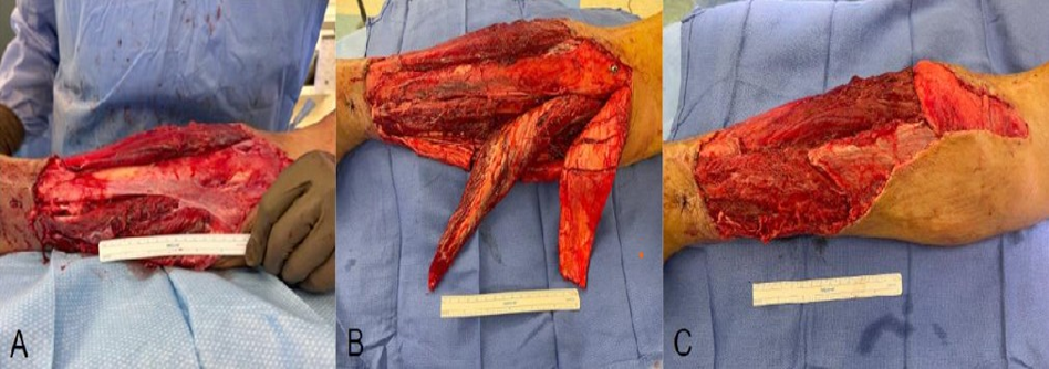
Figure 1: Proximal- and middle-third leg soft tissue defect. A. A large soft tissue defect of the proximal and middle thirds of the lower leg. B. Isolation of the hemisoleus and medial gastrocnemius flaps. C. Transposition of the flaps into the defect.
This technique is used for large proximal- and middle-third defects requiring substantial tissue volume. The hemisoleus flap can be combined with additional regional muscle flaps. The medial gastrocnemius muscle and tibialis anterior muscle can be added to offer coverage for sizable defects. The gastrocnemius muscle can be scored along its fascia to extend its reach. The tibialis anterior, less often needed, can be transposed medially over the tibia while maintaining its proximal and distal attachments, as well as its blood supply and innervation (Figure 1).
Double Hemisoleus Flap
The soleus muscle can be split into medial and lateral hemisoleus flaps, each based on independent vascular pedicles. These segments can be mobilized independently, offering more flexible inset for defects of the middle third of the leg where a single flap may be insufficient. If the medial hemisoleus flap is rotated into a more proximal defect, the distal wound may be amenable to a second, independently dissected flap created by elevating the lateral hemisoleus. The lateral hemisoleus can be rotated to cover the distal aspect of the wound. Separation of the two hemisoleus flaps offers an additional degree of freedom and greater flexibility during inset in comparison to an undivided soleus flap. After elevation of the medial hemisoleus, the posterior aspect of the raphe septum is incised posteriorly along the length of the muscle. The lateral hemisoleus is then similarly dissected off the conjoint tendo-achilles and fascia with the Freer elevator. After the muscle is divided distally, the flap can be rotated into the defect. Additional
dissection is then performed as indicated to facilitate the inset. Care is taken to avoid injury to the proximal blood supply of the peroneal system (Figure 2).
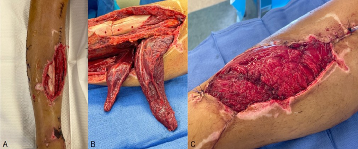
Figure 2: Middle-third leg defect. A. A soft-tissue defect in the middle third of the leg. B. The medial and lateral hemisoleus flaps are isolated. C. Both medial and lateral hemisoleus flaps are rotated into the defect for complete coverage.
Reverse Hemisoleus Flap
The reverse (distally based) hemisoleus flap utilizes perfusion through distal minor perforators, allowing the flap to reach defects in the distal third of the leg. Careful preoperative planning is essential and includes Doppler assessment of distal perforators, supplemented by angiographic evaluation when indicated. Intraoperative Doppler is also used to confirm the presence of adequate distal perforators. The soleus muscle is elevated from proximal to distal, with proximal perforators ligated and distal perforators preserved. Dissection is carried to approximately 6-8 cm proximal to the ankle to maintain reliable distal blood supply. The hemisoleus is then transposed distally into the defect. In cases where a single muscle flap is insufficient to achieve complete coverage, adjunctive reverse muscle flaps (e.g., peroneus brevis) can be utilized to augment coverage. Flap viability is assessed using Doppler, Indocyanine Green (ICG) angiography, and clinical evaluation of bleeding at the flap edges. Nonviable distal muscle is trimmed prior to inset. We recommend staging the skin graft coverage to confirm the viability of the muscle flap (Figure 3).
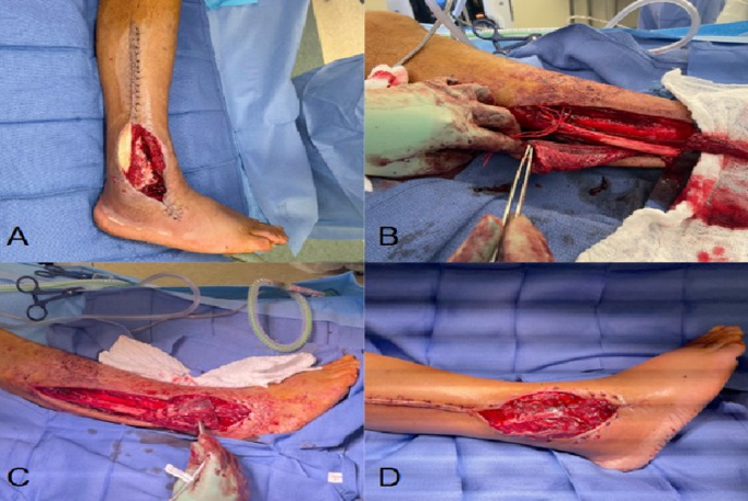
Figure 3: Distal-leg soft-tissue defect. A. A large soft tissue defect in the distal third of the leg. B. Careful dissection of the flap, preserving the distal perforators. C. Inset of the flaps into the defect. D. Closure after distal transposition of the flap into the defect.
Propeller Hemisoleus Flap
The propeller hemisoleus flap is based on a single perforating vessel, allowing the muscle segment to be rotated in a propeller fashion to reach distal defects. By leveraging a reliable perforator and careful dissection, this approach provides an additional option for distal third coverage while maintaining the advantages of local tissue reconstruction. Following exposure of the soleus, meticulous dissection is performed to identify suitable perforators from the posterior tibial vessels. Once identified, the perforator is dissected to free the vessel from any overlying tissue. The hemisoleus segment is divided from proximal and distal attachments and separated along the midline raphe while preserving the perforator pedicle. The muscle segment is then rotated, typically between 90 and 180 degrees, into the defect. Care is taken to avoid torsion or kinking of the pedicle. Perfusion is confirmed with Doppler and bleeding edges prior to definitive inset. This technique allows for increased flexibility in flap positioning and is particularly useful for distal defects when standard reverse-flow options are not feasible (Figure 4).
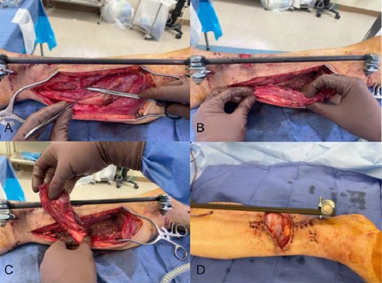
Figure 4: Distal leg soft-tissue defect. A. Identification of suitable perforators. B. Careful dissection of the hemisoleus, preserving the perforator. C. Rotation of the muscle segment into the defect. D. Final closure after the muscle flap was rotated into the defect.
Results
Twelve patients underwent hemisoleus-based reconstruction with complete flap survival in all cases. Each reconstructive approach was selected based on defect location and intraoperative assessment of perforator anatomy. Three patients (25.0%) were current smokers, one (8.3%) was a former smoker, and eight (66.7%) had never smoked. Relevant comorbidities and treatment history included diabetes mellitus in two patients (16.7%), HIV in two (16.7%), and prior cerebrovascular accident, methamphetamine use, and prior radiotherapy in one patient each (8.3%). Hemisoleus-based reconstruction achieved coverage of exposed critical structures in all 12 patients, with no total flap loss documented in the available postoperative records. Minor complications occurred in three patients (25.0%), and one patient (8.3%) experienced a major complication requiring unplanned operative treatment for infection. Minor complications included flapedge necrosis, skin-graft breakdown, and peri-incisional erythema. One patient developed a surgical-site infection requiring operative debridement and was therefore classified as having a major complication (Tables 1,2).
|
Patient |
Age/ Sex |
Diagnosis / Indication |
Defect Location |
Defect Size (cm²) |
Flap Type |
Adjunctive Flap |
STSG |
Complications |
Flap Outcome |
|
#1 |
49/M |
Osteomyelitis with exposed bone/tendon |
Mid-distal tibia |
60 |
Reverse hemisoleus |
Peroneus brevis |
Delayed |
Edge necrosis requiring bedside debridement |
Complete survival |
|
#2 |
49/M |
Chronic tibial osteomyelitis |
Mid tibia |
50 |
Proximally based hemisoleus |
Gastrocnemius |
Yes |
SSI requiring reoperation |
Complete survival |
|
#3 |
40/M |
Traumatic wound with exposed tibia |
Distal tibia |
60 |
Propeller hemisoleus |
None |
Yes |
Skin graft breakdown after noncompliance |
Complete survival |
|
#4 |
48/M |
Traumatic wound with exposed tibia |
Mid tibia |
132 |
Proximally based hemisoleus |
None |
Yes |
Mild periincisional erythema |
Complete survival |
|
#5 |
20/M |
Open tibia/fibula fracture |
Mid-distal tibia |
100 |
Double hemisoleus |
None |
Yes |
None |
Complete survival |
|
#6 |
83/F |
Traumatic wound |
Mid tibia |
600 |
Proximally based hemisoleus |
Gastrocnemius + Tibialis anterior |
Yes |
None |
Complete survival |
|
#7 |
63/M |
Traumatic wound |
Mid tibia |
300 |
Proximally based hemisoleus |
Gastrocnemius + Tibialis anterior |
Yes |
None |
Complete survival |
|
#8 |
51/M |
Open tibia/fibula fracture |
Midproximal tibia |
150 |
Proximally based hemisoleus |
Gastrocnemius |
Yes |
None |
Complete survival |
|
#9 |
57/M |
Traumatic wound |
Mid tibia |
450 |
Proximally based hemisoleus |
Gastrocnemius |
Yes |
None |
Complete survival |
|
#10 |
64/M |
Leiomyosarcoma resection defect |
Mid tibia |
78 |
Proximally based hemisoleus |
Tibialis anterior |
Yes |
None |
Complete survival |
|
#11 |
74/F |
Necrotizing fasciitis s/p debridement |
Midproximal tibia |
200 |
Proximally based hemisoleus |
Gastrocnemius |
Delayed |
None |
Complete survival |
|
#12 |
31/M |
Open tibia/fibula fracture |
Mid tibia |
200 |
Proximally based hemisoleus |
None |
Yes |
None |
Complete survival |
Table 1: Patient Characteristics, Operative Details, and Outcomes.
Abbreviations: STSG, split-thickness skin graft; SSI, surgical-site infection; s/p, status post; M, male; F, female.
|
Technique |
Indication |
Vascular Basis |
Main Advantage |
|
Proximally based +/- adjunct |
Large proximal/mid defects |
Proximal posterior tibial perforators |
Increased area of coverage |
|
Reverse |
Distal third defects |
Distal minor perforators of posterior tibial or peroneal systems |
Coverage of distal third defects |
|
Propeller |
Distal third defects |
Single isolated perforator arising from the posterior tibial or peroneal vessels. |
Allows distal reach of the muscle flap. Preserves vascular supply to the distal limb. |
|
Double hemisoleus |
Large/complex defects |
Independent medial/lateral pedicles off the posterior tibial and peroneal systems. |
Increased area of coverage. Provides flexibility of inset. |
Table 2: Characteristics of Hemisoleus-Based Reconstructive Approaches.
Discussion
This case series demonstrates that hemisoleus-based muscle flaps can provide reliable and versatile coverage for complex softtissue defects of the middle and distal lower extremity. Complete flap survival was achieved in all 12 patients despite substantial variation in defect size, etiology, and reconstructive technique. Three patients experienced minor wound complications and one required reoperation for surgical-site infection; however, none of these events resulted in partial or total flap loss. The complete flap survival observed in this series is consistent with the high rates of flap survival reported in prior hemisoleus series. One patient developed recurrent osteomyelitis after being lost to follow-up and required hardware removal and debridement. The flaps themselves remained viable. No additional muscle flap was required. Other studies have found similar success with their hemisoleus reconstructions. Pu reported successful wound healing in all 14 patients undergoing proximally based medial hemisoleus reconstruction, although one patient developed minor distal flap necrosis requiring debridement and flap advancement [3]. Similarly, Schierle et al. achieved successful coverage and wound healing in all 17 patients treated with distally based hemisoleus flaps, with one case of distal tip necrosis [8]. Our experience with combined regional muscle flaps also compares favorably with that of Ong et al., who reported survival of all 10 combined gastrocnemius–hemisoleus flaps, with two minor complications [13]. While these prior studies generally evaluated individual flap designs, this series extends these observations across a broader range of hemisoleus-based reconstructive techniques and clinical indications. The principal advantage of the hemisoleus flap is the versatility afforded by its anatomy and vascular supply. The soleus is a Mathes-Nahai type II muscle, supplied by a dominant proximal pedicle with additional minor segmental pedicles. This vascular anatomy, together with the ability to longitudinally divide and selectively mobilize the muscle, permits several distinct reconstructive configurations. Flap design can therefore be tailored to defect location, size, depth, and available vascular anatomy.
Several cases in this series required modification of the initial reconstructive plan based on intraoperative findings, highlighting the hemisoleus flap’s adaptability as an option for wound coverage and the value of maintaining multiple regional reconstructive options when approaching complex lower extremity defects. Although free tissue transfer remains an important standard for many complex lower extremity defects, local and regional muscle flaps continue to offer meaningful advantages. Hemisoleus-based reconstruction avoids microsurgical anastomosis, preserves recipient vessels, and reduces dependence on microsurgical resources [6, 13]. Thornton et al. reported longer operative times and higher costs for free muscle flap reconstruction compared with local flap reconstruction in complex lower extremity wound coverage [15]. These advantages may be particularly relevant in rural or resource-limited settings, where access to microsurgical expertise, specialized equipment, and intensive postoperative flap monitoring may be limited. Importantly, local flap reconstruction remains an effective component of contemporary lower extremity reconstruction even where microsurgical capabilities are available. Györi et al. compared local and free flap reconstruction of distal lower extremity defects and demonstrated an overall limb preservation rate of 94.1%, supporting the continued role of local tissue reconstruction alongside free tissue transfer [16]. Regional hemisoleus-based reconstruction may therefore represent a particularly useful option when defect characteristics permit local coverage or when microsurgical reconstruction is unavailable or impractical. This does not diminish the role of free tissue transfer for larger or more complex defects, but underscores the importance of maintaining regional flap techniques within the reconstructive armamentarium. Traditional lower extremity reconstructive algorithms have generally favored the soleus for middle-third defects and free tissue transfer for distal-third wounds. However, improved understanding of local vascular anatomy has challenged these conventional boundaries. Song and Pu emphasized selection of local versus free tissue transfer according to patient and defect characteristics rather than location alone [17]. For distal hemisoleus reconstruction specifically, Schierle et al. demonstrated reliable coverage in 17 patients using an angiosome-based design that emphasized preservation of the distal perforator territory, with only one case of distal tip necrosis [8]. These principles are reflected in our approach, in which flap design is determined not solely by defect location but also by the vascular anatomy available to support the proposed reconstruction. Assessment of perforator anatomy may therefore expand local reconstructive options in appropriately selected distal defects while identifying cases in which an alternative regional flap or free tissue transfer is more appropriate. Saleh et al. described a “reversed flow hemisoleus propeller muscle flap,” reporting reliable coverage of distal leg defects [9]. While the terminology overlaps with our propeller flap configuration, the two techniques are fundamentally distinct. In the technique described in our series, the propeller flap is based on a single perforating vessel arising from the posterior tibial bundle, consistent with the accepted definition of a propeller flap. In comparison, the technique described by Saleh et al. relies on a reversed-flow posterior tibial pedicle and does not meet the criteria for previously described perforator-based propeller flaps [18,19]. This key distinction between techniques allows the posterior tibial vessels to be preserved, maintaining antegrade flow to the distal limb, which is an important consideration in patients with trauma, diabetes, or vascular disease. Additionally, it keeps the posterior tibial vessel available as a recipient for later free tissue transfers or bypasses. Although we did not formally measure flap reach in our series, we acknowledge that a reversed-flow propeller flap design may afford a greater arc of rotation and distal reach compared to our perforator-based propeller configuration. This study has several limitations. It is retrospective, includes a small number of patients, and reflects the experience of a single center. Follow-up duration was variable, and standardized functional outcome measures were not available for all patients. In addition, flap selection was individualized rather than protocolized, limiting direct comparison among flap configurations. Nevertheless, this series illustrates the application of multiple hemisoleus-based strategies across a heterogeneous group of lower extremity defects and provides a basis for further study of technique-specific indications and outcomes.
Conclusion
Hemisoleus-based reconstruction provided soft-tissue coverage in this 12-patient series using several flap configurations, alone or in combination with other regional muscle flaps. No total flap loss was observed, although wound complications occurred and one patient required operative treatment for infection. These findings demonstrate that the hemisoleus-based flaps are a versatile option for wound coverage and support their consideration for selected lower extremity defects, with flap design guided by defect characteristics and available vascular anatomy. The hemisoleus muscle flap can provide durable limb-salvage coverage while preserving the practical advantages of local tissue reconstruction and represents an important component of the contemporary lower extremity reconstructive armamentarium. Larger studies with standardized follow-up and functional assessment are needed to better define technique-specific outcomes and donor-site morbidity.
Acknowledgements and Funding: There are no acknowledgements. This study received no specific funding.
Ethical Considerations: This study was conducted in accordance with applicable institutional and ethical standards. Institutional review board approval was obtained prior to data collection at the Queen’s Medical Center, IRB RA-2025-029. Given the retrospective nature of the study, the requirement for informed consent was waived by the institutional review board.
Conflict of Interest: The authors declare no conflicts of interest.
References
- Tobin GR (1985) Hemisoleus and reversed hemisoleus flaps. Plast Reconstr Surg 76: 87-96.
- Fayman MS, Orak F, Hugo B, Berson SD (1987) The distally based split soleus muscle flap. Br J Plast Surg 40: 20-26.
- Pu LLQ (2006) Soft-tissue coverage of an open tibial wound in the junction of the middle and distal thirds of the leg with the medial hemisoleus muscle flap. Ann Plast Surg 56: 639-643.
- Pu LLQ (2006) The reversed medial hemisoleus muscle flap and its role in reconstruction of an open tibial wound in the lower third of the leg. Ann Plast Surg 56: 59-63.
- Pu LLQ (2008) Further experience with the medial hemisoleus muscle flap for soft-tissue coverage of a tibial wound in the distal third of the leg. Plast Reconstr Surg 121: 2024-2028.
- Souza Filho MVP, Teixeira JCEO, Castro OC (2011) Reversed hemisoleus flap for wound coverage in the distal third of the leg. Rev. Bras. Cir. Plást 26: 710-713.
- Fisal AA, Abdel-Hamid Romeih M, Younes LM, El-Rosasy MA, Rodríguez P, et al. (2020) Distally Based Medial Hemisoleus Muscle Flap for Wound Coverage in the Distal Third of the Leg. Clin Podiatr Med Surg 37: 631-647.
- Schierle CF, Rawlani V, Galiano RD, Kim JYS, Dumanian GA (2009) Improving outcomes of the distally based hemisoleus flap: principles of angiosomes in flap design. Plast Reconstr Surg 123: 1748-1754.
- Saleh DMG, Shaker AA, Alrefahi M, Estawrow MA (2023) The Reversed Flow Hemisoleus Propeller Muscle Flap. Ann Plast Surg 90: 76-81.
- Thornton BP, Pu LLQ (2004) Reconstruction of an extensive tibial softtissue defect with multiple local muscle flaps for limb salvage when free-tissue transfer was not an option. Eur J Plast Surg 27: 217-221.
- Hyodo I, Nakayama B, Takahashi M, Toriyama K, Kamei Y, et al. (2004) The gastrocnemius with soleus bi-muscle flap. Br J Plast Surg 57: 77-82.
- Pu LLQ (2010) Soft-tissue coverage of an extensive mid-tibial wound with the combined medial gastrocnemius and medial hemisoleus muscle flaps: the role of local muscle flaps revisited. J Plast Reconstr Aesthet Surg 63: e605-e610.
- Ong SW, Gan LP, Chia DSY (2019) The double muscle gastrocnemius– soleus flap in resurfacing large lower limb defects: Modifications and outcomes. J Orthop 17: 13-16.
- Jitprapaikulsarn S, Benjawongsathien K, Patamamongkonchai C, Gromprasit A, Thremthakanpon W (2021) Combined medial gastrocnemius and hemisoleus flap: A reproducible alternative for open tibial fractures complicated with large or double soft tissue defects. Eur J Orthop Surg Traumatol 31: 413-420.
- Thornton BP, Rosenblum WJ, Pu LLQ (2005) Reconstruction of limited soft-tissue defect with open tibial fracture in the distal third of the leg: a cost and outcome study. Ann Plast Surg 54: 276-280.
- Györi E, Fast A, Resch A, Rath T, Radtke C (2022) Reconstruction of traumatic and non-traumatic lower extremity defects with local or free flaps. Eur Surg 54: 44-49.
- Song P, Pu LLQ (2018) The soleus muscle flap: an overview of its clinical applications for lower extremity reconstruction. Ann Plast Surg 81: S109-S116.
- Mehrotra S (2007) Perforator-plus flaps: a new concept in traditional flap design. Plast Reconstr Surg 119: 590-598.
- Yajima H, Tamai S, Ishida H, Fukui A (1995) Partial soleus muscle island flap transfer using minor pedicles from the posterior tibial vessels. Plast Reconstr Surg 96: 1162-1168.