Taiceer Abdulwahab*, Neha Muralidharan, Sattar Alshryda, Suresh Annamalai
Department of Orthopaedic Surgery, King’s College Hospital London, Dubai Hills, Dubai
*Corresponding Author: Taiceer Abdulwahab, Department of Orthopaedic Surgery, King’s College Hospital London, Dubai Hills, Dubai
Received Date: 01 June 2026
Accepted Date: 06 June 2026
Published Date: 08 June 2026
Citation: Abdulwahab T, Muralidharan N, Alshryda S, AnnamalaiS (2026) Successful Management of Stage I Congenital Knee Dislocation using a Pavlik Harness as a Novel Treatment Method: A Case Report. J Surg 11:11641DOI: 10.29011/2575-9760.011641
Abstract
Congenital Knee Dislocation (CKD) is a rare musculoskeletal anomaly characterised by hyperextension of the knee with varying degrees of anterior tibial displacement. Early recognition and prompt conservative management are critical, particularly in milder forms. The gold-standard treatment is above-knee serial casting, which is the current recommended treatment in literature [1,2] present a case of Stage I CKD successfully treated with initial manipulation and Pavlik harness, resulting in full functional recovery. This case highlights the efficacy of a novel method of early non-operative treatment, which reinforces current principles in the management of mild congenital knee deformities.
Keywords: CKD: Congenital Knee Dislocation: Pavlik Harness
Introduction
Congenital Knee Dislocation (CKD) is a rare condition with an estimated incidence of approximately 1 in 100,000 live births. It is characterised by hyperextension deformity at the knee, often accompanied by anterior subluxation or dislocation of the tibia relative to the femur. CKD is considered as a part of ‘packaging disorders’, frequently associated with developmental dysplasia of the hip (50%), syndromic conditions such as arthrogryposis multiplex congenita and Larsen syndrome, although it may occur in isolation. [3] The classification system most used is that described by dividing CKD into three stages graded by passive range of flexion and radiologic appearance and can guide treatment (Table 1, Figure 1)
|
Grade |
Radiology |
Range of passive flexion |
|
I |
Knee Recurvatum |
>90° |
|
II |
Subluxation |
30°-90° |
|
III |
Dislocation |
<30° |
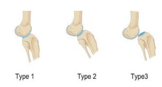
Figure 1: Tarek Classification of CKD [4].
Management depends on severity at presentation and timing of diagnosis. Early conservative treatment remains the cornerstone for Stage I deformities, with excellent reported outcomes. We present a case demonstrating successful correction using Pavlik harness as ana lternative to traditional serial casting.
Case Presentation
A full-term neonate was referred on first day of life with left knee hyperextension deformity noted at birth. This was the second child for the mother, with a previous healthy child and no knee deformity. The pregnancy had been uncomplicated, with no history of oligohydramnios or intrauterine constraint. There was no family history of musculoskeletal or syndromic conditions. On examination, left knee demonstrated marked hyperextension of approximate 20 degrees beyond neutral. Passive flexion was possible to 70 degrees without resistance. Hip examination was normal. Bilaterally, there was no thigh crease and no leg length discrepancy, with Ortolani and Barlow negative and full range of hip abduction. The feet were plantigrade, and there were no associated deformities, spinal or dysmorphic features (Figure 2).
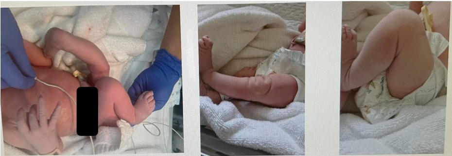
Figure 2: Clinical appearance of left knee recurvatum 20 degrees achieving 70 degrees of flexion at birth presentation.
Ultrasound knee confirmed hyperextension at the knee joint without evidence of subluxation or dislocation, easily reducible into full flexion with a congruent knee joint. Radiologically there was no evidence of anterior tibial subluxation, and the joint remained congruent on gentle manipulation, consistent with Stage I congenital knee dislocation. Ultrasound of the hip showed left hip Graf type IIA immature hip with an alpha angle of 58 degrees, dynamically stable, and more than 2/3rds of the femoral head within the socket. The right hip was unremarkable with alpha angle >60degrees and Graf type I.
Results
A Pavlik harness was applied with the hips in approximately 90 degrees of flexion and the knees flexed to the maximum achievable angle without undue force (approximately 100 degrees initially) as shown in (Figure 3). The patient was reviewed at weekly intervals under careful clinical supervision. After a total of four weeks, the deformity had fully corrected, with the knee achieving full flexion and extension within normal physiological limits. The harness was discontinued, the knee was clinically stable, with no recurvatum noted on active or passive range of motion, indicating the knee was stable. There were no concerns regarding the hip. The patient was transitioned to physiotherapy focusing on gentle range-of-motion exercises.
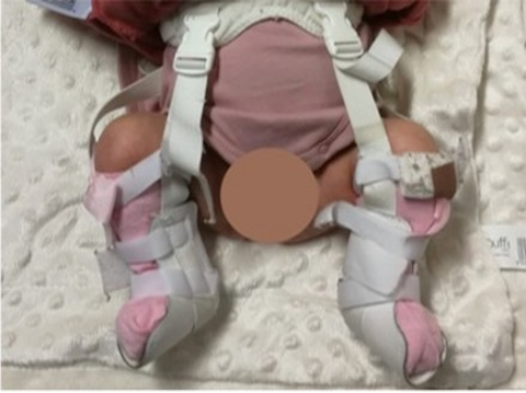
Figure 3: Child in Pavlik Harness. Photo credit:Leuzinger-Dias C, Fonseca M, Cabral J, Alves C (2024) Congenital dislocation of the knee: a surprise in the delivery room. J Neonatol Clin Pediatr 11: 132.
Discussion
The underlying pathology in CKD includes quadriceps contracture, capsular tightness, and varying degrees of ligamentous imbalance. In Stage I cases, these changes are mild and highly responsive to stretching and gradual correction along the viscoelastic principles of stress relaxation. The Pavlik harness has been used for hip dysplasia and femoral fractures, but this is a novel application for CKD. Given the early presentation and classification as CKD Tarek Stage I, the standard management would have been gentle manipulation with serial casting. Although this is the ‘gold standard’, this has issues in itself, casting requires application training similar to that of Posetti method for congenital club foot, weekly long-leg plaster changes, associated plaster complications, added costs of application, and weekly changes can cause distress to parents. The Pavlik harness is more baby friendly and tolerated than applying an above knee plaster, and taking advantage of the anterior knee strap for knee flexion is advantageous if needed to treat hip dysplasia concomitantly, however treatment depends on clinical and radiological findings. The management of CKD is guided primarily by the severity of deformity and timing of intervention. Stage I deformities, as in this case, have an excellent prognosis when treated early with conservative measures. More studies to show Pavlik harness as treatment of CKD Stage I and II are required.
Conclusion
This case illustrates that Stage I congenital knee dislocation can be effectively managed with early Pavlik harnessing, even in the presence of mild hip dysplasia without the need for serial weekly castings resulting in excellent functional outcomes. Prompt diagnosis is essential to avoid surgical intervention. Failure of conservative treatment is usually associated with delayed diagnosis or syndromic associations (e.g. arthrogryposis). Importantly, overtreatment should be avoided. Aggressive manipulation risks physeal injury or iatrogenic instability. Early conservative management should remain the first-line treatment in mild cases, with close follow-up to ensure sustained correction and normal development.
Ethical Guidelines
Verbal consent was obtained from the parents for publication of this case report and accompanying images. All procedures performed were in accordance with the ethical standards of the institutional and/or national research committee and with the principles of the Declaration of Helsinki.
Conflict of Interest: No conflicts of interest that may influence the results or interpretations of the manuscript.
References
- Curtis BH, Fisher RL (1970) Congenital dislocation of the knee. J Bone Joint Surg Am. 52: 539-550.
- Jacobsen K, Vopalecky F (1985) Congenital dislocation of the knee. Acta Orthop Scand. 56: 1-7.
- Oetgen ME, Walick KS, Tulchin K, Karol LA (2010) Functional results of congenital knee dislocation. J Pediatr Orthop. 30: 216-223.
- Abdelaziz TH, Samir S (2011) Congenital knee dislocation: a protocol for management based on degree of knee flexion. J Child Orthop. 5: 143-149.